Microarray Spotting & Hybridization
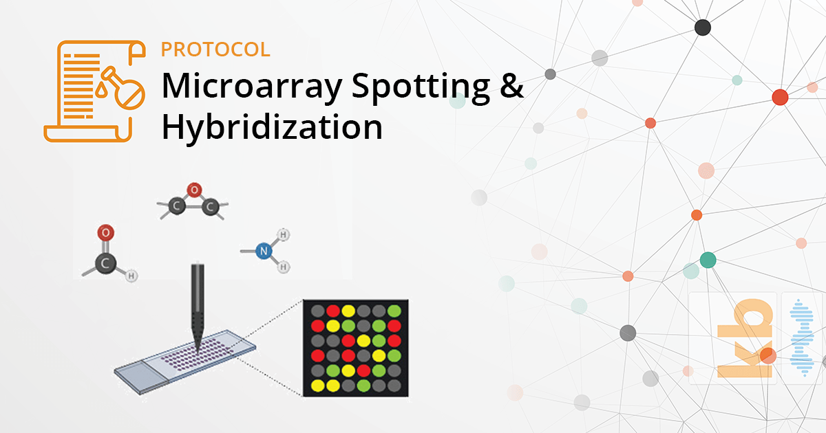
Microarray Spotting & Hybridization
Chemical coating of slides is a widely used practice for optimizing microarray methods and analysis. The use of various chemicals and compounds has traditionally proven to significantly improve technical features such as; molecule retention and post-hybridization signal amplification, for covalent attachment of unmodified and modified fragments. One of the most critical stages in a microarrays workflow is the spotting of slides. Spotting techniques can strongly impact the quality of hybridization along with the array analysis. The use of slides coated with chemical or functional compounds requires special handling during these steps. The following series of protocols demonstrate the proper handling of the three most common coated-slides; Epoxy, Aldehyde, & Amine-based.
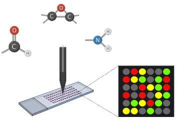
Table of Contents
- Epoxy Coated Arraying Slides
- Aldehyde Arraying Slides
- Amine Arraying Slides
- Aldehyde Spotting Solution
- Amine Spotting Solution
Epoxy Coated Arraying Slides
Introduction
Epoxy slides are epoxy-coated arraying slides that provide a uniform substrate for the creation of high-quality nucleic acid microarrays. These slides provide robust immobilization chemistry as the epoxy coating reacts with amino, hydroxy, or thiol-containing biomolecules and forms a covalent bond with the activated glass surface. Therefore, efficient covalent and directed binding of molecules, such as oligonucleotides and/or PCR products is possible. Unmodified DNA can be directly spotted on these slides. These slides can also be used for covalent binding of proteins and cells. The epoxy substrate supports low endogenous fluorescence and provides high signal-to-noise ratio. The epoxy bond is probably the most robust attachment chemistry available to the microarray scientist today.
Protocol
Array Printing
Use an appropriate spotting solution to array your DNA targets on the slides. Resuspend your DNA targets at a concentration of 0.1-1.0 µg/µl in the spotting solution for arraying. Coupling between DNA and the epoxy-coated surface should be complete within 15 minutes of spotting.
Slides should be stored unprocessed. They should be processed, as needed, for hybridization. The slide processing protocol is provided below:
Slide Processing
- Wash slide twice in 2X SSC / 0.1% SDS at room temperature for 2 minutes.
- Denature in dH20 at 95-100°C for 2 minutes. Allow to cool for 30 seconds.
- Dry the slides by placing in a slide dryer and centrifuging for 1 minute at 100 x g (1000rpm).
Hybridization of the processed slide can be carried out as per protocol below:
Hybridization and Imaging
- Hybridize the slide with a labeled probe in genHYB microarray hybridization buffer, using a hybridization chamber or similar, sealed humid chamber.
- Hybridize the slide according to the protocol for the genHYB microarray hybridization buffer.
- Transfer the slide to a slide washer and wash according to the genHYB hybridization protocol.
- Place the slide in a slide dryer and centrifuge at 100 x g (1000 rpm) for 1 minute.
- Visualize the array by scanning with an appropriate fluorochrome channel on a scanner.
Aldehyde Arraying Slides
Introduction
Aldehyde slides are aldehyde-coated slides designed for microarray production. The slides carry functional aldehyde groups for the covalent attachment of reactive amine-containing molecules. They covalently bind targets to the glass surface via the Schiff base aldehyde-amine chemistry. Lysine residues of proteins or primary amines in DNA bases can participate in Schiff base formation. Uniform coating and high binding capacity allow consistent and reproducible arraying of amine rich or modified target molecules including PCR products, oligonucleotides, and amine rich protein.
Note: Most aldehyde coated slides are not compatible for spotting DNA samples in DMSO, or standard Amine Spotting Solution.
Protocol
Array Printing
Array your targets onto the slides using industry-standard Aldehyde Microarray Spotting Solution or another appropriate spotting solution, such as 3X SSC). Resuspend your DNA targets at a concentration of 0.1-1.0 µg/µl in the appropriate spotting solution for arraying.
Following arraying, leave the slides for a minimum of 4 hours in a constant humidity environment. Slides should be processed as per protocol below before hybridization.
Slide Processing
- Wash slide twice in 0.2% SDS at room temperature for 2 minutes.
- Wash slide twice in dH20 at room temperature for 2 minutes.
- Wash in dH20 at 95-100°C for 2 minutes. Allow to cool for 30 seconds.
- Wash for 5 minutes in Sodium borohydride mix at room temperature (1g Sodium borohydride dissolved in 300ml 1x PBS, and 100ml 100% ethanol).
- Wash 3 times in 0.2% SDS at room temperature for 1 minute.
- Rinse twice in dH20 for 2-3 seconds at room temperature.
- Dry the slides by placing in a slide dryer and centrifuging for 1 minute at 100 x g (1000rpm) for 1 minute.
- Store slides in the dark at room temperature. At this point, the processed slides can be stored for several months.
Hybridization of the processed slide can be carried out as per protocol below:
Hybridization and Imaging
- To hybridize your slide, react the slide with a labeled probe in genHYB microarray hybridization buffer, using a hybridization chamber or similar sealed humid chamber.
- Hybridize the slide according to protocol for the hybridization buffer.
- Transfer the slide to a slide washer and wash according to the genHYB hybridization protocol.
- Place the slide in a slide dryer and centrifuge at 100 x g (1000 rpm) for 1 minute. Visualize the array by scanning with an appropriate fluorochrome channel on a scanner.
Amine Arraying Slides
Introduction
Amine slides are slides designed for microarray production. The slides bind your targets through amine-reactive groups on the slide surface. Ionic bonds are formed between the surface amine groups and the targets positively charged groups. This is supplemented by the UV treatment of the spotted slide which gives additional non-specific covalent binding of the target to the slide. They are particularly suited to spotting unmodified PCR products. This has the advantage of not requiring the user to amino-modify all their targets i.e. amino-modified primers need not be used for amplification of the targets.
Protocol
Array Printing
Array your targets onto the slides using industry standard Amine Microarray Spotting Solution or another appropriate spotting solution, such as 3X SSC). Resuspend your DNA targets at a concentration of 0.1-1.0 µg/µl in the appropriate spotting solution.
UV crosslink the targets to the surface of the slide (approximately 120 mJoules per cm2.) Alternatively, bake the slides at 80°C for 1-2 hours. Process slides as per protocol below:
Slide Processing
- Wash slide twice in 0.1% SDS at room temperature for 5 minutes.
- Treat the slides in blocking solution (0.75g succinic anhydride dissolved in 125ml 1-methy-2-pyrrolidinone then 125 ml 0.2M boric acid pH 8 added) at room temperature for 20 minutes.
- Wash 3 times in dH20 at room temperature for 1 minute.
- Denature the double-stranded DNA by washing in dH20 at 95-100°C for 2 minutes.
- Dry the slides by placing in a slide dryer and centrifuging for 1 minute at 100 x g (1000 rpm) for 1 minute.
- Store slides in the dark at room temperature. At this point the processed slides can be stored for several months.
Hybridization and Imaging
- To hybridize your slide, react the slide with a labeled probe in genHYB microarray hybridization buffer using a hybridization chamber or similar sealed humid chamber.
- Hybridize the slide according to protocol for the hybridization buffer.
- Transfer the slide to a slide washer and wash according to the genHYB hybridization protocol.
- Place the slide in a slide dryer and centrifuge at 100 x g (1000 rpm) for 1 minute. Visualize the array by scanning with an appropriate fluorochrome channel on a scanner.
Aldehyde Spotting Solution
Introduction
Aldehyde Microarray Spotting Solution can be used to generate consistent and robust spotting of your DNA targets on microarray slides with surface aldehyde chemistry (aldehyde arraying slides). Its unique constituents stabilize the DNA during the target printing and drying process, which gives a more uniform spot size and shape, thus improving the overall quality of the array.
Protocol
- Re-suspend your DNA samples in an appropriate volume of dH20 to give you a concentration of 20-100 pmole/µl for oligonucleotides or 0.4-2.0 µg/µl for cDNAs. Alternatively, if you have performed a clean-up on your PCR samples, calculate the DNA concentration in the eluate and adjust to a concentration of 20-100 pmole/ml for oligonucleotides or 0.4-2.0 µg/µl for cDNAs.
- Add an equal volume of Microarray Spotting solution. Mix by pipetting up and down several times.
- Using an appropriate volume for each well, transfer the sample to a 96-well or 384-well microplate.
- Print the samples onto suitable slides using a robotic microarrayer.
- Process the slides for hybridization, according to the protocol for the chosen substrate type.
Amine Spotting Solution
Introduction
Amine Microarray Spotting Solution can be used to generate consistent and robust spotting of your DNA targets on microarray slides with surface amine chemistry (amine arraying slides). Its unique constituents stabilize the DNA during the microarray fabrication and drying, which gives a more uniform spot size and shape, thus improving the overall quality of the array. This spotting solution is recommended for use with cDNA and oligonucleotide spotting on amine slides.
Protocol
- Re-suspend your DNA samples in an appropriate volume of dH20 to give you a concentration of 20-100 pmole/µl for oligonucleotides or 0.4-2.0 µg/µl for cDNAs. Alternatively, if you have performed a clean-up on your PCR samples, calculate the DNA concentration in the eluate and adjust to a concentration of 20-100 pmole/ml for oligonucleotides or 0.4-2.0 µg/µl for cDNAs.
- Add an equal volume of Microarray Spotting solution. Mix by pipetting up and down several times.
- Using an appropriate volume for each well, transfer the sample to a 96-well or 384-well microplate.
- Print the samples onto suitable slides using a robotic microarrayer.
- Process the slides for hybridization, according to the protocol for the chosen substrate type.